Imaging using incoherent light

Physicists demonstrate new method for resolving nanostructures
Physicists at the Friedrich Alexander University Erlangen-Nürnberg (FAU), the University of Hamburg and the research facility DESY have succeeded for the first time in revealing tiny structures using an imaging method that relies on the usual diffraction of light, but which does not require the scattered light to be coherent. With conventional imaging methods using the diffraction of coherent light, scientists have to go to considerable lengths to ensure the coherence of the radiation, i.e. the electromagnetic waves must remain in phase during the scattering process. The new method uses incoherent light instead. The process, which has now been demonstrated for the first time using soft X-ray radiation, stands to revolutionise the methods of diffractive optics and has been published in the journal Nature Physics.
For over 100 years, the diffraction of coherent X-rays has been used to determine the structure of crystals and molecules. The method relies on diffraction and interference, properties that are displayed by all waves: light waves, which are made up of photons, are deflected by the atoms in a crystal and interfere with each other. If a sufficient number of such photons are measured by a detector, they produce a characteristic diffraction image, which can be used to calculate the arrangement of the atoms within the crystal. However, this requires the waves to be scattered coherently. When the photons are not scattered coherently, in other words there is no fixed phase relationship between the incident and diffracted wave, conventional X-ray imaging methods are no longer able to supply information about the arrangement of the atoms.
This effect limits the use of coherent diffractive X-ray imaging. “In the case of X-rays, incoherent scattering usually predominates, for example when fluorescent light is produced by photon absorption followed by photon emission,” explains Anton Classen from FAU, who is the principal author of a recent paper in the journal Physical Review Letters where the method was first presented theoretically. “This creates diffuse background radiation, which has until now been assumed to be unusable for imaging, instead reducing the fidelity of the imaging using coherent methods.”
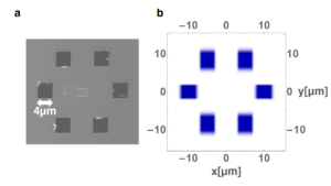
Now, scientists have used the very same incoherent radiation to record a structure shaped like a benzene ring with the help of diffuse scattered light in the soft X-ray range at DESY’s free-electron laser.
“Our work provides the basis for a fundamentally new approach to X-ray imaging, because our experiment has demonstrated that incoherently scattered photons can be used to reconstruct complex structures at short wavelengths,” explains Raimund Schneider of FAU, the principal author of the paper on the experimental findings.
The fundamental technique used is not entirely new. Robert Hanbury Brown and Richard Q. Twiss already used incoherent light back in 1956 to determine the diameter of stars (HBT effect). In the years that followed, however, the technique received little attention in terms of its capacity to measure very small structures. “Although it was perfectly possible to determine the diameter of a single object, such as a star, it was by no means clear that the method could be applied in microscopic structural analysis too, and certainly not to unravel complex geometries using a multitude of light sources in two or three dimensions,” notes Joachim von Zanthier, a professor at FAU. “Another key aspect is that, while the HBT experiment only uses the second-order intensity correlation, we determined intensity correlations all the way up to the fifth degree, and then used these to reconstruct the arrangement of the scattering centres. This means that the structural information produced by our method is split into smaller components and can therefore be determined more easily and with higher precision.”

Free-electron lasers like FLASH or the European X-ray Free-Electron Laser (European XFEL) have an extremely high brilliance and produce ultra-short X-ray pulses, offering ideal conditions for this new approach to structural analysis, according to Wilfried Wurth, who is the scientific director of FLASH and a professor at the University of Hamburg.
The novel method has a further crucial advantage. “The smaller the structures you want to examine, the larger the proportion of incoherently scattered light,” explains the co-author Ralf Röhlsberger who works at DESY and is also a professor at Hamburg University. “Whereas coherent imaging has problems with the increasing intensity, our method benefits from it.” The method therefore has the potential to fundamentally improve structural analyses in biology and medicine.
Nature Physics (DOI: 10.1038/nphys4301)
Physical Review Letters (DOI: 10.1103/PhysRevLett.119.053401)
The method is also described in a „Brennpunktartikel“ of the December issue of the german „Physik Journal“
Further informationen:
Prof. Dr. Joachim von Zanthier
Phone: +49 9131/85-27603
joachim.vonzanthier@physik.uni-erlangen.de